Using AI to control energy for indoor agriculture
30 September 2024
Published online 13 March 2014
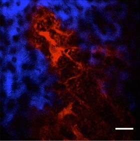
A bespoke imaging technique has uncovered a surprising oxygen distribution in bone marrow, helping researchers better understand the microenvironment of stem cells.
The researchers, led by a team based at Harvard Medical School and including Walid Zaher of King Saud University, Saudi Arabia, used an oxygen-sensitive phosphorescent dye to directly measure the partial pressure of oxygen in the bone marrow of live mice. Their results, published in Nature, confirmed earlier measurements showing that bone marrow contains relatively little oxygen.
The non-invasive, high-resolution imaging technique also revealed a "unique hypoxic landscape", where the amount of oxygen varied with the distribution and types of blood vessels in the marrow. To the team's surprise, the oxygen gradient was the opposite of what had been predicted. They also discovered that the hypoxic landscape varied in response to stresses such as irradiation or chemotherapy.
"We kept getting results that were unexpected," says Charles Lin, an associate professor at Harvard Medical School and senior author of the study. "We had to change our thinking to understand our own data."
These findings challenge the idea that low oxygen levels are an important component of the stem cell microenvironment. Other factors may be at play, since some likely stem cell niche locations have relatively high oxygen concentrations.
doi:10.1038/nmiddleeast.2014.63
30 September 2024
23 September 2024
11 September 2024
Sign-up to receive our e-alert update every two weeks to keep up with everything new on the portal.
Sign up for e-alerts
Stay connected: