Using AI to control energy for indoor agriculture
30 September 2024
Published online 30 January 2017
Newly discovered direction and distance nerve cells in the brains of Egyptian fruit bats could help scientists understand how the brain conducts complex computations and why Alzheimer’s patients are often spatially disoriented.
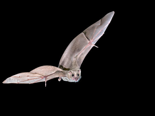
Nachum Ulanovsky and colleagues from the Weizmann Institute of Science in Israel wirelessly measured brain nerve activity while trained bats navigated to a specific goal. They found 58 neurons in the brain’s hippocampus— known for its role in memory and navigation—that fired off according to the bats’ movement in relation to the goal’s direction. Another 49 cells fired off as the bats got closer to the goal.
Researchers have known for decades that the brain contains specific cells—called place, grid and head-direction cells—that represent an animal’s own location and orientation. But they didn’t know how navigational goals were encoded in the brain.
“Studying navigation at the neuronal level is one of the best models we can use to learn how the brain performs behaviourally-relevant computations, such as distance, direction and speed,” says Ayelet Sarel, one of the study’s co-authors. “We can learn general concepts about how the brain performs computations from its navigation system,” she adds.
Ulanovsky’s team implanted tiny electrodes in the brains of Egyptian fruit bats, which wirelessly recorded the activity of 309 neurons located in a region of the hippocampus called CA1. The bats were trained to fly in complex trajectories in a large room and land on a single landing site.
The presence of ‘goal-direction’ and ‘goal-distance’ cells in the hippocampus suggests that animals navigate by representing their goal with a vector: a line that represents the direction and distance between the animal’s and the goal’s location. “Although this idea has existed for years, based purely on behavioural evidence, our study is the first to show neurons that could support goal-directed navigation using vectors,” says Sarel.
Understanding the brain’s navigation system could improve understanding of conditions, such as Alzheimer’s disease, in which the hippocampus is damaged and patients’ orientation in space is affected, Sarel says.
“Despite decades of research, cells with similar activity patterns have not been identified in the rodent hippocampus, which might suggest key differences in the cognitive mechanisms supporting goal-directed navigation in rats and bats," says Daniel Bush of University College London's Institute of Cognitive Neuroscience.
"These findings will allow us to generate new theoretical models of mammalian navigation, better constrained by an understanding of how the brain encodes the relative location of important goals.”
The study has raised many more questions that the team hopes to investigate. They would like to further understand how animals navigate in the wild, how they represent more than one goal at a given time, and how these representations are modified in response to environmental changes.
doi:10.1038/nmiddleeast.2017.26
30 September 2024
23 September 2024
11 September 2024
Sign-up to receive our e-alert update every two weeks to keep up with everything new on the portal.
Sign up for e-alerts
Stay connected: