Using AI to control energy for indoor agriculture
30 September 2024
Published online 27 November 2019
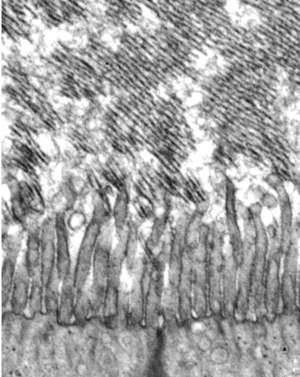
An inner ear membrane is anchored to the cell surface during development and grows one layer at a time.
Sungjin Park
The prevailing hypothesis suggests that the components of the tectorial membrane are released into the network of molecules surrounding cells, known as the extracellular matrix, and then begin to self-assemble. But Sungjin Park of the University of Utah and his team weren’t convinced. “We reasoned that the tectorial membrane is too complex to be organized solely by an extracellular assembly process,” he explains. “We looked for an alternative model and observed that the border between the cell and the extracellular matrix plays a critical role in tectorial membrane morphogenesis.”
His team, including Ali Almishaal, who is currently affiliated with Saudi Arabia’s University of Hail, hypothesized that TECTA, a protein anchored to the cells on which the tectorial membrane develops, might guide tectorial membrane development by preventing its components from diffusing into the extracellular space. To test this, they engineered a version of TECTA lacking the anchor that tethers it to the cell. They found that mice with the mutated TECTA protein formed a severely disorganized tectorial membrane that was detached from its normal location.
Next, the researchers made a version of TECTA that was attached to the membrane by a transmembrane domain rather than the usual anchor domain. This led to the accumulation of tectorial membrane components on the cell surface, but because TECTA release was impaired, the tectorial membrane didn't develop into a multi-layered structure.
Based on these findings, the researchers suggest that the tectorial membrane forms via a process similar to 3D printing. TECTA on the cell surface serves as an organizer for tectorial membrane components; once a layer is formed, TECTA is released and the next layer is ‘printed’ underneath it, eventually resulting in a multi-layered architecture.
These findings not only clarify how the tectorial membrane develops but could also shed light on other structures that develop in the extracellular matrix. “The proposed 3D printing model will provide a novel insight into our understanding of extracellular matrix morphogenesis process during development and tissue repair,” says Park.
doi:10.1038/nmiddleeast.2019.156
Kim, D-K. et al. The release of surface-anchored α-tectorin, an apical extracellular matrix protein, mediates tectorial membrane organization. Sci. Adv. 5, eaay6300 (2019).
30 September 2024
23 September 2024
11 September 2024
05 April 2018
20 June 2016
27 September 2012
Sign-up to receive our e-alert update every two weeks to keep up with everything new on the portal.
Sign up for e-alerts
Stay connected: