The Missing Kingdom: Why Fungi Must Be Central to Conservation Strategy
28 December 2025
Published online 1 June 2022
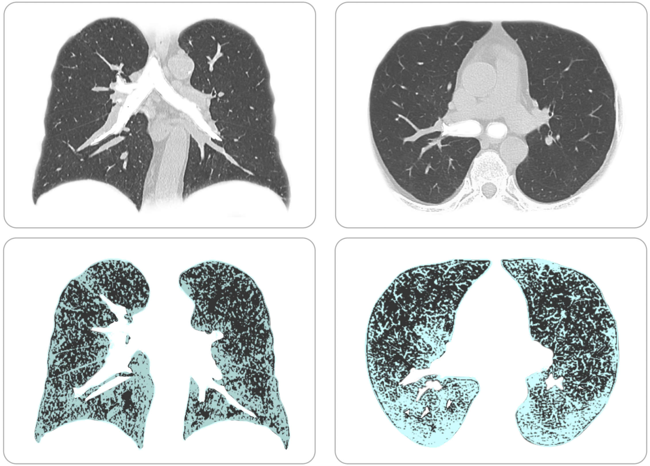
Scientists use AI to enhance the resolution of lung abnormalities in CT scans.
Longxi Zhou
Enlarge image
The Deep-Lung Parenchyma-Enhancing (DLPE) approach calculates the optimal signal window for observing normally invisible lung lesions in CT scans. Existing methods, including computer-based ones, fail to detect this damage, which appears in CT scans as ground-glass opacities, which describes a scratched-looking surface, and lung scars.
Normally, to view lung lesions, radiologists need to reduce the CT signal range to what is called the lung window. But the signals coming from sub-visual lesions and healthy background tissue when viewed within this window are not very distinct from each other. Also, blood vessels and airways have strong CT signals, further hampering the ability to see the lung lesions. “DLPE calculates the scan-optimal window for observing the lesions in individual patients, enhancing them dozens of times and making them visible,” explains PhD computer science student, Longxi Zhou from King Abdullah University of Science and Technology (KAUST) in Saudi Arabia.
The research team, from Saudi Arabia and China, trained their model using thousands of CT scans from healthy people and people who were hospitalized with COVID-19, who remain ill. Without DLPE, they were only able to detect a small volume of lesions, whereas DLPE allowed them to find a much larger volume.
“We also found that the method dramatically improved the prediction of key clinical metrics, like the gas exchange ratio in the lungs, by assessing the volume of the lesions,” says KAUST computer scientist, and computational biologist, Xin Gao.
The researchers say that such knowledge will help clinicians assess the extent of lung damage, and determine the desired therapy.
The method could also be used to detect mild lung tissue scarring and decipher minute features of various lung diseases like pneumonia, tuberculosis and lung cancers.
doi:10.1038/nmiddleeast.2022.29
Zhou, L. et al. An interpretable deep learning workflow for discovering subvisual abnormalities in CT scans of COVID-19 inpatients and survivors. Nat. Mach. Intell. https://doi.org/10.1038/s42256-022-00483-7 (2022).
28 December 2025
24 December 2025
24 December 2025
01 December 2020
19 June 2020
18 March 2020
Sign-up to receive our e-alert update every two weeks to keep up with everything new on the portal.
Sign up for e-alerts
Stay connected: