Understanding longevity: From gene sequences to social inequity
03 April 2025
Published online 4 May 2023
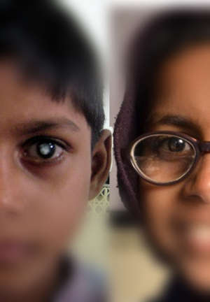
The brain’s ability to interpret visual information remains plastic throughout adolescence, with implications for treating congenital blindness with eye surgery.
Bas Rokers & Pawan Sinha, 2023
“There has long been debate whether, and to what extent, the human visual system remains plastic as the brain develops,” says Bas Rokers at New York University Abu Dhabi, UAE, who led the study alongside a colleague, Caterina Pedersini, in collaboration with scientists in India. “The current consensus is that, after a critical period has closed, at around seven years of age, the visual system becomes resistant to change. We wanted to examine this conjecture.”
The team studied 19 patients aged 7 to 16 years from India, who had dense cataracts in both eyes. The researchers followed the patients through cataract surgery with the aim of differentiating between age- and surgery-related changes in the patients’ neural pathways and their visual capabilities following surgery.
“Because the young people in our study grew up blind, their visual system did not develop in the normal way,” explains Pedersini. “Our study provided a unique chance to identify neural changes in the visual system specifically following surgery.”
The team asked each participant to undertake visual acuity and face perception tasks in the days and weeks following surgery. The patients also underwent regular MRI scans to examine longitudinal white matter changes in the brain. The team then compared these results with brain image databases from the human connectome project (HCP), which contain information on brain development throughout the entire childhood developmental trajectory.
Eye surgery induced significant white matter plasticity in specific ‘late-visual’ neural pathways. These pathways showed enhanced structural integrity after surgery, with improved connections between the occipital region at the back of the brain, which processes visual information from the eyes, and key interpretative brain regions. The patients’ ability to recognise faces, and their visual perception abilities, improved dramatically, enhancing their quality of life.
“We identified the precise neural pathways that underpin this dramatic improvement,” says Rokers. “These visual perception changes were independent of normal maturation changes, and suggest that white matter in the visual system remains plastic throughout adolescence.”
The team hopes their study will reinvigorate discussion around the value of eye surgery in cases of congenital blindness, regardless of the child’s age. The evidence for residual neural plasticity might also trigger the search for behavioural or pharmacological interventions that restore or improve vision, notes Rokers.
“Our next step is to explore the neural deficits underlying more moderate visual impairment, such as lazy eye,” says Pedersini. “If these visual impairments can be attributed to abnormalities in the same pathways, this may open new treatment avenues.”
doi:10.1038/nmiddleeast.2023.42
Pedersini, C.A. et al. White matter plasticity following cataract surgery in congenitally blind patients. PNAS 120,19 (2023).
03 April 2025
18 March 2025
30 September 2024
20 March 2019
13 January 2020
21 March 2021
Sign-up to receive our e-alert update every two weeks to keep up with everything new on the portal.
Sign up for e-alerts
Stay connected: