Using AI to control energy for indoor agriculture
30 September 2024
Published online 12 May 2023
A breakthrough imaging method reveals the complex structural details of zeolite crystals and could help scientists optimize these valuable materials.
Yu Han & Hui Zhang, 2023
Enlarge image
“Transmission electron microscopy (TEM) is a potent tool for revealing structural details of materials, which is crucial for understanding their properties and behaviours,” says Yu Han at King Abdullah University of Science and Technology (KAUST), in Saudi Arabia, who led the study with colleagues, and researchers across China, to develop a novel way of imaging zeolites.
“Investigating zeolites using TEM is challenging and requires special low-dose techniques, because such materials are sensitive to the electron beam, which can actually damage the zeolite’s crystal structure during imaging,” Han continues. “Using TEM for zeolites has further limitations, including thickness-limited resolution, lack of depth resolution, and the need for precise focusing.”
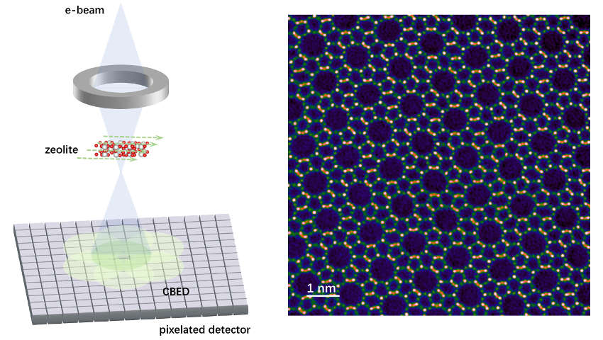
Han and the team developed a ptychography imaging method based on four-dimensional scanning TEM (4D-STEM). Ptychography is a computational method that processes the interference patterns that have been scattered by an object – in this case, the zeolite crystals. Generating multiple images of the object from all different angles above a pixelated detector base made it easier to layer up all the images precisely, creating a robust, highly detailed picture.
“Our technique produced record-breaking resolution for zeolite imaging across a broad range of specimen thicknesses up to 40 nanometres, which conventional TEM techniques cannot achieve,” says Han. “This nanometre-level depth resolution enabled us to create 3D structural images of the zeolites. Our method is more efficient than existing techniques because it eliminates the need for precise focusing prior to imaging.”
The resulting images are so detailed that they highlight the individual oxygen atoms and the three-dimensional distribution of oxygen vacancies within the crystal framework. The images also precisely determine the orientation and behaviour of adsorbed molecules. This imaging breakthrough could help refine and optimize the structure of synthetic zeolites for industrial applications.
“We aim to broaden our research to include other zeolite systems for more complex structural characterizations, focusing on locating dopant atoms within zeolites,” says Han. “This requires enhanced depth resolution and reconstruction accuracy. Furthermore, we will explore 4D-STEM ptychography’s potential to examine extremely sensitive materials including metal-organic frameworks and hybrid perovskites.”
doi:10.1038/nmiddleeast.2023.51
Zhang, H. et al. Three-dimensional inhomogeneity of zeolite structure and composition revealed by electron ptychography. Science 380,6645 (2023).
30 September 2024
23 September 2024
11 September 2024
08 August 2022
09 April 2022
05 October 2020
Sign-up to receive our e-alert update every two weeks to keep up with everything new on the portal.
Sign up for e-alerts
Stay connected: